

A pelvic radiograph of a 5-year-old girl shows a large lytic lesion in the left iliac bone (arrows).
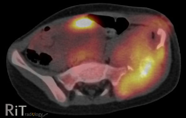

An axial FDG PET/CT image shows high metabolic activity of the mass involving the left iliac bone with soft tissue component and bone destruction. A coronal T2W MR image reveals an extensive soft tissue mass with necrotic areas and involvement of the adjacent musculature.
Differential Diagnosis
- Metastatic neuroblastoma. Given her age at five years old, this needs to be in differentials
- Ewing's sarcoma
- Telangiectatic osteosarcoma
- Osteomyelitis. Great mimics of aggressive-looking bone tumor. Symptoms may overlap with round-cell tumor, including fever
This case: Ewing's sarcoma by tissue diagnosis (+ve PAS and vimentin).
Facts: Ewing's Sarcoma
- Malignant round-cell tumors of the bone with neural cell origin
- Tumors of children and young adults, most between 10-20 years old. Less than 2% occur in children less than 5 years old
- Most common sites = femur >> pelvis
- Pelvic Ewing's -- bad prognosis because there is no anatomic barrier to tumor spread, close proximity to viscera and neurovascular bundles, prone to recur
Reference:
Bhagat S, Sharma H, Pillai DS, Jane MJ. Pelvic Ewing's sarcoma: a review from Swedish Bone Tumour Registry. J Orthop Surg 2008;16:333-8


No comments:
Post a Comment